| Health / Health News |
Structural states of a brain receptor revealed
NIH | SEPTEMBER 2, 2014
Scientists determined the detailed structure and movement of the glutamate receptor, a protein in nerve cells involved in learning, memory, and several diseases.
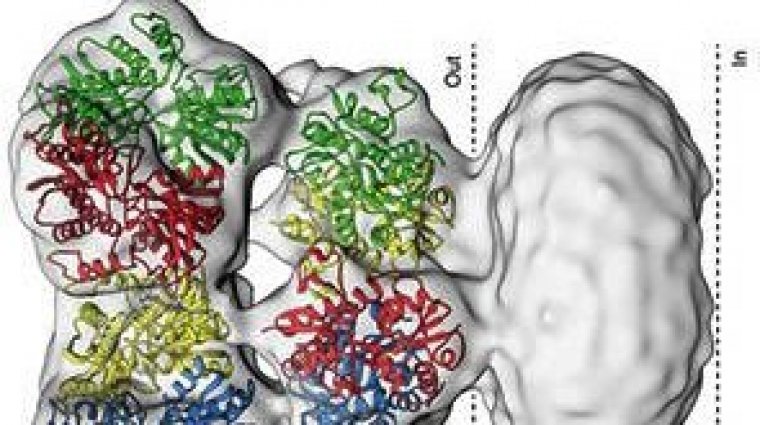
Structure of the glutamate receptor, showing the part where glutamate binds outside the cell and the part that spans the cell membrane (between the dotted lines). Image by the authors.
Cells have receptors on their surface that serve as gatekeepers to transmit signals between the outside and inside. Nerve cells contain glutamate receptors that span the cell membrane. When glutamate binds the receptor on the outside, the receptor changes shape and opens within milliseconds. This allows small substances known as ions to enter the cell.
The process is crucial for communication between nerve cells and plays a role in brain development, learning, and memory. Problems with glutamate receptor function are thought to be involved in many disorders, including autism, schizophrenia, depression, Parkinson’s disease, and some types of cancer.
A team led by Dr. Sriram Subramaniam of NIH’s National Cancer Institute (NCI) set out to determine the structure of the glutamate receptor as it transitions from its resting state to open to desensitized (reduced response to glutamate) states.
A structure of one glutamate receptor variant in its resting state had previously been determined by using X-ray crystallography. This technique requires a protein to first be crystallized into a fixed 3-D shape. However, capturing the protein’s multiple states in a crystal is challenging, and could potentially trap subtly misfolded or incorrect structures.
The team used an imaging technique called cryo-electron microscopy. This method allowed them to capture high-resolution snapshots of receptors in several transitional stages.
The group analyzed detailed images of different types of glutamate receptors and captured the receptors in different states. They determined that the transition of the protein from the resting to open states involved a corkscrew-like rotational motion within the protein. The shift from open to desensitized states required much larger rearrangements.
The scientists note that while several technical hurdles remain, the technique will allow them to analyze the structures of many other proteins, as well as study receptor-drug interactions.
YOU MAY ALSO LIKE


