| News / Science News |
New Findings Reveal Surprising Role of the Cerebellum in Reward and Social Behaviors
Researchers found a direct neural connection from the cerebellum to the ventral tegmental area (VTA) of the brain, which is an area long known to been involved in reward processing and encoding.
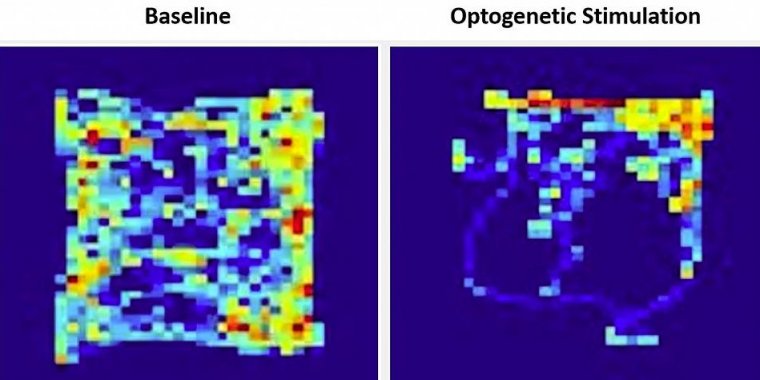
Heatmaps showing the amount of time a mouse spent in locations of the open field chamber at baseline (left) and during optogenetic stimulation of the cerebellar input to the VTA (right). The “reward quadrant” is located in the upper right section of the field. Photo: Albert Einstein College of Medicine
The cerebellum plays a well-recognized role in the coordination and regulation of motor activity. However, research has also suggested that this brain area contributes to a host of non-motor functions.
For example, abnormalities in the cerebellum have been linked to autism, schizophrenia, and substance use disorders, and brain activation in the cerebellum has been linked to motivation, social and emotional behaviors, and reward learning, each of which can be disrupted in psychiatric disorders.
To examine this, the researchers used a technique called optogenetics, in which the neurons of animals are genetically modified, so they can be controlled using pulses of light.
The researchers found that activating the cerebellar neurons led to increased activation in the VTA of mice, indicating a working connection between these two brain structures.
Mice showed a strong preference for spending time in the reward quadrant, freely choosing to spend more than 70 percent of their time in this area. In addition, the researchers found that mice were willing to work for activation in this brain area and to spend time in conditions they would usually not prefer (light vs. darker areas) to receive this activation.
Together, the findings suggest that activation of the cerebellar projections to the VTA is rewarding for mice and that the cerebellum plays a role in reward-related behaviors.
To examine whether the inputs from the cerebellum into the VTA impacted social behaviors, the researchers tested mice using a three-chamber social task in which the mice could choose to spend time in a chamber with another mouse (the social chamber), in an empty central chamber, or in a chamber containing a non-social object.
At baseline, mice preferred to be in the social chamber, but after researchers inactivated the cerebellar projections into the VTA, the mice no longer showed this preference.
In addition, continuous silencing of the cerebellar-VTA pathway was found to completely prevent the expression of social preference behavior in the mice, findings which indicate that inputs from the cerebellum into the VTA are necessary for social preference behavior in mice.
The results of this study suggest a potentially major—and previously unrecognized—role for the cerebellum in the creation and control of reward and social preference behaviors.
Although there is much left to explore, the identification of this direct neural pathway may help explain the role of this circuit in disorders that involve reward-related and social-processing systems, such as addiction, autism, and schizophrenia, and may point to future targets for intervention and symptom management. (National Institutes of Health)
YOU MAY ALSO LIKE


