| Health / Health News |
Visual activity regenerates neural connections between eye and brain
NIH | JULY 20, 2016
A study in mice shows for the first time that high-contrast visual stimulation can help damaged retinal neurons regrow optic nerve fibers, otherwise known as retinal ganglion cell axons.
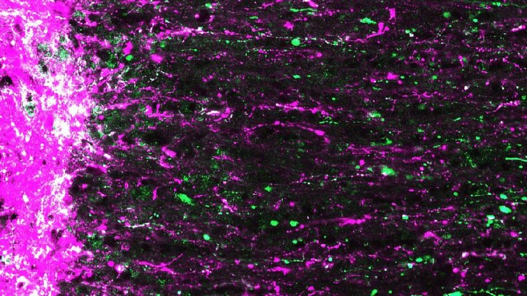
Regenerating mouse retinal ganglion cell axons (magenta and green) extending from site of optic nerve injury (left). ![]()
In combination with chemically induced neural stimulation, axons grew further than in strategies tried previously. Treated mice partially regained visual function. The study also demonstrates that adult regenerated central nervous system (CNS) axons are capable of navigating to correct targets in the brain.
The researchers induced optic nerve damage in mice using forceps to crush the optic nerve of one eye just behind the eyeball. The mice were then placed in a chamber several hours a day for three weeks where they viewed high-contrast images—essentially changing patterns of black lines. The mice had modest but significant axonal regrowth compared to control mice that did not receive the high-contrast visual stimulation.
Prior work by the scientists showed that increasing activity of protein called mTOR promoted optic nerve regeneration. And so they wondered if combining visual stimulation with increased mTOR activity might have a synergistic effect.
Two weeks prior to nerve crush, the scientists used gene therapy to cause the retinal ganglion cells to overexpress mTOR. Optic nerve crush was performed and mice were exposed to high-contrast visual stimulation daily. After three weeks, the scientists saw more extensive regeneration, with axons growing through the optic nerve as far as the optic chiasm, a distance from the eye of about 6 millimeters.
Encouraged by these results, the researchers again increased mTOR activity but then forced mice to use the treated eye during visual stimulation by suturing shut the good eye. This combined approach of increasing mTOR activity with intense visual stimulation promoted regeneration down the full length of the optic nerve and into various visual centers of the brain.
We saw the most remarkable growth when we closed the good eye, forcing the mice to look through the injured eye,” said Andrew Huberman, Ph.D., associate professor, Stanford University School of Medicine’s department of neurobiology. In three weeks, the axons grew as much as 12 millimeters, a rate about 500 times faster than untreated CNS axons.
For future therapies that preserve optic nerve axons, Huberman envisions the development of filters for virtual reality video games, television programs, or eyeglasses designed to deliver regeneration-inducing visual stimulation.
YOU MAY ALSO LIKE


